

Mammography - A Lifesaving Tool for Early Breast Cancer Detection
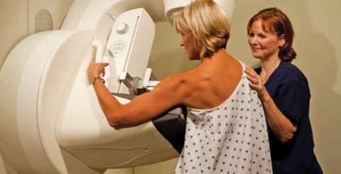
Breast cancer is one of the most common cancers affecting women worldwide. Early diagnosis plays a vital role in improving treatment outcomes and survival rates. Among the most reliable screening methods available today, mammography remains the gold standard for detecting breast abnormalities, often before they become noticeable through physical examination. Mammography is a specialized imaging technique that uses low-dose X-rays to examine breast tissue. It helps identify breast cancer at an early stage and also assists in diagnosing various non-cancerous breast conditions. Regular mammographic screening has significantly reduced breast cancer-related mortality by enabling timely diagnosis and treatment.
What is Mammography?
Mammography is a medical imaging procedure that uses low-energy X-rays to create detailed images of the breast, known as mammograms. These images allow healthcare professionals to detect abnormal changes within the breast tissue, including tumors, microcalcifications, cysts, and other structural abnormalities that may not be detectable during a physical examination. During the procedure, each breast is gently compressed between two plates attached to the mammography machine. Although this compression may cause temporary discomfort, it is necessary to obtain high-quality images while minimizing radiation exposure.
Modern mammography systems are available in two forms
- 2D Digital Mammography (Traditional Mammography): Produces two-dimensional X-ray images of the breast from different angles.
- 3D Digital Breast Tomosynthesis (DBT): Captures multiple images from different angles and reconstructs them into a three-dimensional view, allowing better visualization of overlapping breast tissues. This technology has improved the detection of small cancers while reducing false-positive results, particularly in women with dense breast tissue.
Types of Mammography
Mammography is generally categorized into two main types based on its purpose.
1. Screening Mammograph
Screening mammography is performed in women who have no signs or symptoms of breast disease. Its primary objective is to identify breast cancer before it becomes clinically apparent. Routine screening is recommended for women within the appropriate age group and those with average risk, as advised by their healthcare provider. Detecting cancer at an early stage often allows for less invasive treatment and significantly improves survival rates.
2. Diagnostic Mammography
Diagnostic mammography is recommended when a woman experiences symptoms such as:
- A breast lump
- Breast pain
- Nipple discharge
- Skin thickening or dimpling
- Changes in breast size or shape
- An abnormal finding on a screening mammogram
Unlike routine screening, diagnostic mammography involves additional views and more detailed imaging to evaluate suspicious areas. It helps determine whether the abnormality is benign or requires further investigation through ultrasound, MRI, or biopsy.
Why is Mammography Important?
Mammography has transformed breast cancer screening by enabling healthcare providers to detect disease before symptoms develop. Its importance extends beyond cancer diagnosis.
1. Early Detection of Breast Cancer
One of the greatest advantages of mammography is its ability to identify tiny tumors and microcalcifications long before they can be felt during a breast examination. Early detection increases treatment options and greatly improves the likelihood of successful recovery.
2. Reduced Breast Cancer Mortality
Numerous long-term clinical studies have demonstrated that regular mammographic screening significantly lowers breast cancer-related deaths. Women diagnosed during the early stages generally have better treatment outcomes and higher survival rates.
3. Detection of Benign Breast Conditions
Not every breast abnormality indicates cancer. Mammography also helps diagnose several non-cancerous conditions, including:
- Breast cysts
- Fibrocystic breast changes
- Benign calcifications
- Fat necrosis
- Fibroadenomas
Identifying these conditions helps physicians determine appropriate monitoring or treatment while avoiding unnecessary concern.
Supports Treatment Planning
When breast cancer is diagnosed, mammography provides valuable information regarding the tumor's size, location, and extent. This information assists healthcare professionals in planning surgery, radiation therapy, chemotherapy, or other treatment approaches.
Benefits of Mammography
Mammography offers several important advantages:
- Detects breast cancer before symptoms appear.
- Improves survival by facilitating early diagnosis.
- Provides a quick and minimally invasive imaging procedure.
- Requires no surgical incision or anesthesia.
- Helps distinguish between benign and suspicious breast abnormalities.
- Assists physicians in monitoring previously identified breast conditions.
- Offers improved accuracy with modern digital and 3D imaging technologies.
Limitations of Mammography
Although mammography is highly effective, it is not without limitations.
1. False-Positive Results
Occasionally, mammograms identify abnormalities that ultimately prove to be non-cancerous. These findings may require additional imaging or biopsy, causing temporary anxiety for patients.
2. False-Negative Results
Mammography may occasionally miss breast cancers, particularly in women with dense breast tissue where tumors can be difficult to distinguish from normal tissue.
3. Temporary Discomfort
Breast compression during imaging may cause brief discomfort or mild pain. However, the procedure usually lasts only a few minutes, and the discomfort quickly resolves afterward.
4. Low Radiation Exposure
Mammography uses a very small amount of ionizing radiation. The radiation dose is considered extremely low, and for most women, the benefits of early cancer detection far outweigh the minimal associated risk.
Cost of Mammography
The cost of mammography varies depending on the healthcare facility, city, and technology used.
In India, approximate costs are:
- Standard 2D Mammography: ₹1,500–₹2,500
- Digital 3D Mammography (Tomosynthesis): ₹5,000–₹8,000
Prices may differ across hospitals and diagnostic centers.
Frequently Asked Questions
1.Why is breast compression necessary during mammography?
Compression spreads the breast tissue evenly, reduces image blurring, lowers radiation exposure, and improves image quality, enabling radiologists to detect even very small abnormalities.
2. What is digital mammography?
Digital mammography uses electronic detectors instead of traditional film to capture breast images. These digital images can be enlarged, stored electronically, and reviewed more efficiently, improving diagnostic accuracy.
3. When should women start getting mammograms?
Screening recommendations vary among medical organizations and should be individualized based on age, family history, and overall breast cancer risk. For women at average risk, many healthcare providers recommend beginning regular screening around 40 years of age, while some guidelines recommend annual screening between 45 and 54 years, followed by screening every two years after 55 years. Women with a strong family history, genetic predisposition, or other high-risk factors may require earlier and more frequent screening. It is always best to discuss the most appropriate screening schedule with a qualified healthcare provider.
Conclusion
Mammography remains one of the most valuable tools for the early detection of breast cancer. By identifying abnormalities before symptoms develop, it allows timely intervention, expands treatment options, and significantly improves survival rates. Although mammography has certain limitations, its benefits far outweigh its risks for most women. Regular screening, combined with breast self-awareness, routine clinical examinations, and a healthy lifestyle, forms a comprehensive approach to maintaining breast health. Consulting a healthcare professional about the appropriate screening schedule based on individual risk factors is an important step toward early detection and better long-term outcomes.
Similar Articles

Is it possible becoming a mom after successful breast cancer therapy? It's not easy, but it is possible. Breast cancer hits younger women in many cases. Many of those affected are still childless when diagnosing cancer and would like to have a child after successful breast cancer therapy.

So, you’ve been diagnosed with breast cancer. Now, when you can finally speak, help from your husband and your breast cancer specialist can hardly be overestimated.