

7 Early Detection Strategies That Reduce Breast Cancer Mortality
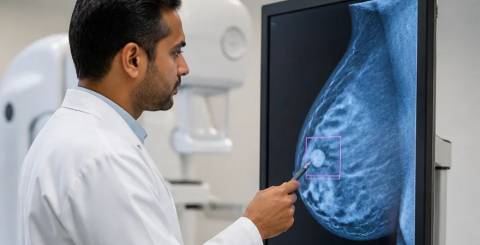
Breast cancer remains one of the most commonly diagnosed cancers among women worldwide, yet survival rates have improved dramatically over the past three decades. That progress stems largely from detection methods that identify tumors before they spread beyond the surrounding tissue. When caught at stage one, the five-year survival rate exceeds 99 percent, compared with roughly 29 percent for stage four diagnoses. The seven strategies below cover which screening approaches actually work, when to begin them, and how combining several methods turns a life-threatening illness into a treatable condition.
1. Assess Your Personal Risk Before Choosing a Screening Schedule
Medical organizations publish baseline recommendations, but blanket guidance does not account for the factors that make one woman's risk very different from another's. A proper risk assessment considers family history, genetic mutations such as BRCA1 and BRCA2, previous biopsies showing atypical hyperplasia, radiation exposure to the chest before age 30, and tissue density.
Women with a lifetime risk above 20 percent typically qualify for supplemental MRI alongside standard imaging. Those carrying known genetic mutations may begin surveillance in their twenties or thirties, often combining several imaging methods to overcome the limits of any single test. Knowing where you sit on that spectrum is the first decision that shapes every other one.
2. Begin Mammography at the Right Age and Keep the Interval
Most guidelines suggest women at average risk start annual or biennial mammography between ages 40 and 50, depending on the organization, and continue for as long as they remain in good health. Mammography is still the only screening tool proven through randomized trials to reduce breast cancer mortality in average-risk populations, which is why it anchors almost every national program.
Consistency matters more than any single appointment. A missed interval is the most common reason a tumor is found later than it needed to be. Research on mammography for early breast cancer detection explains why population-level programs continue to treat it as their foundation.
3. Ask Whether 3D Tomosynthesis Is Available
Digital mammography and the newer 3D tomosynthesis create layered images that reduce tissue overlap, which matters most in dense breasts where tumors hide behind glandular structures. Studies show tomosynthesis raises detection rates by roughly 20 to 65 percent compared with standard 2D imaging, while simultaneously cutting the false positives that lead to unnecessary biopsies.
Not every facility offers it, and not every woman needs it. Asking the question before booking costs nothing, and the answer can meaningfully change what the radiologist is able to see.
4. Add Supplemental Imaging When Breast Tissue Is Dense
Dense tissue appears white on a mammogram, and so do tumors, which makes small malignancies genuinely difficult to visualize. Nearly half of all women in screening age groups have heterogeneously or extremely dense breasts.
For these women, supplemental ultrasound detects cancers that mammography misses, though it raises false positives too. Magnetic resonance imaging offers the highest sensitivity of any method but carries higher costs, longer scan times, and a false positive rate that makes it impractical as a universal tool. Contrast-enhanced mammography and molecular breast imaging are emerging as alternatives when cost or claustrophobia rules out MRI.
5. Build the Habit of Monthly Breast Awareness
Imaging finds tumors too small to feel, but awareness still catches what falls between appointments. Women who examine themselves regularly become familiar with the normal texture and architecture of their own tissue, which makes it far easier to notice a new lump, skin dimpling, nipple retraction, or unusual discharge.
Randomized trials have not shown that formal self-examination reduces mortality on its own, yet roughly 25 percent of breast cancers are discovered by women themselves between scheduled screenings. Technique matters: examine in a systematic pattern, use flat fingers rather than pinching, and check during the same phase of the menstrual cycle each month. A clear walkthrough of breast self examination makes the method easy to repeat correctly.
6. Combine Clinical Examination With Imaging
A clinical breast examination performed by a trained provider adds a layer that neither imaging nor self-checks replace. A clinician can assess subtle skin changes, evaluate lymph nodes, and identify palpable masses that justify imaging even in women not yet eligible for routine mammography.
Its value as a standalone screening tool remains debated, and reasonably so. As one component of a combined strategy built on patient awareness, clinical assessment, and appropriate imaging, it closes gaps that the other two leave open.
7. Weigh the Benefits Against the Harms Honestly
Screening involves trade-offs worth understanding before committing to a schedule. Mammography finds many cancers early, but it also identifies slow-growing tumors that might never have caused symptoms, which leads to overdiagnosis and overtreatment. False positives create real anxiety: women screened annually from 40 to 49 have roughly a 60 percent chance of at least one false positive recall across that decade.
Radiation exposure is another consideration, although the dose from modern digital units is low and the prevention benefit clearly outweighs the risk after age 40. Younger women face higher relative radiation risk, which is precisely why recommended start ages differ. Reviewing current breast cancer screening guidelines helps weigh these factors against a personal risk profile and personal values.
Making the Strategy Stick
The future of breast cancer screening lies in risk-stratified approaches that match intensity and frequency to individual likelihood of disease. Predictive models combining genetic factors, tissue density, reproductive history, and lifestyle already estimate personal risk with growing accuracy. Artificial intelligence tools that read mammograms show promise for improving detection and easing radiologist workload, while liquid biopsy tests that detect circulating tumor DNA may eventually enable blood-based screening.
Cost and access remain practical barriers everywhere. Even within advanced healthcare systems, screening rates vary by income, insurance coverage, and distance from a clinic. Programs that reduce financial barriers, deploy mobile mammography units, and offer multilingual patient education close those gaps faster than any new machine.
Whatever the technology, the most effective program remains the one women actually complete. Shorter waits, prompt results, and supportive clinical environments improve adherence more reliably than equipment upgrades. Combining evidence-based imaging, clinical expertise, personal awareness, and honest risk assessment gives any woman her best chance of catching breast cancer while treatment still works well.
Similar Articles

Breast cancer is one of the most common cancers affecting women worldwide. Early diagnosis plays a vital role in improving treatment outcomes and survival rates.

Is it possible becoming a mom after successful breast cancer therapy? It's not easy, but it is possible. Breast cancer hits younger women in many cases. Many of those affected are still childless when diagnosing cancer and would like to have a child after successful breast cancer therapy.

So, you’ve been diagnosed with breast cancer. Now, when you can finally speak, help from your husband and your breast cancer specialist can hardly be overestimated.